Skip to content

American Society of Pediatric Nephrology (ASPN)

Member Login
  • About
    • Council Members
    • Membership Services
    • Affiliates
    • Awards & Grants
    • Newsletters
    • Constitution & Bylaws
    • ASPN Past Presidents
    • In Memoriam
      • H. William Schnaper Tribute Page
    • Contact Us
    • Privacy Policy
    • Strategic Plan
  • Education and Meetings
    • Upcoming Events
    • Alnylam Webinar: More Than Just A Kidney Stone
    • 8th Annual Multidisciplinary Symposium Registration
    • 2020 Virtual Summer Sessions
    • ASPN Meetings at PAS
    • ASPN Meetings at ASN
    • Other Meetings
    • ASPN Leadership Development Program
    • Board Review Course
    • Past Meetings
    • Webinars
    • Outside Sources
  • Trainee Info
    • pFeNA
    • Fellows Educational Grants
    • Annual Meeting Awards
    • Careers in Nephrology
    • JELF Advocacy Scholars Program
  • Committees
    • Audit Committee
    • Awards
    • Certification
    • Clinical Affairs & Practice Management
    • Communications
    • Global Health
    • Member Education
    • Membership
    • Nominating
    • pFeNA
    • Program
    • Public Policy
      • KCP – Quality & Payment
    • Research
    • Therapeutics Development
    • Workforce
  • Marketplace
    • Employment Ads
    • Careers in Nephrology
  • ASPN Foundation
    • Donate
    • Programs
    • History Project

Acute interstitial nephritis

» 2020 » June » 10

Acute interstitial nephritis

10 Jun 2020

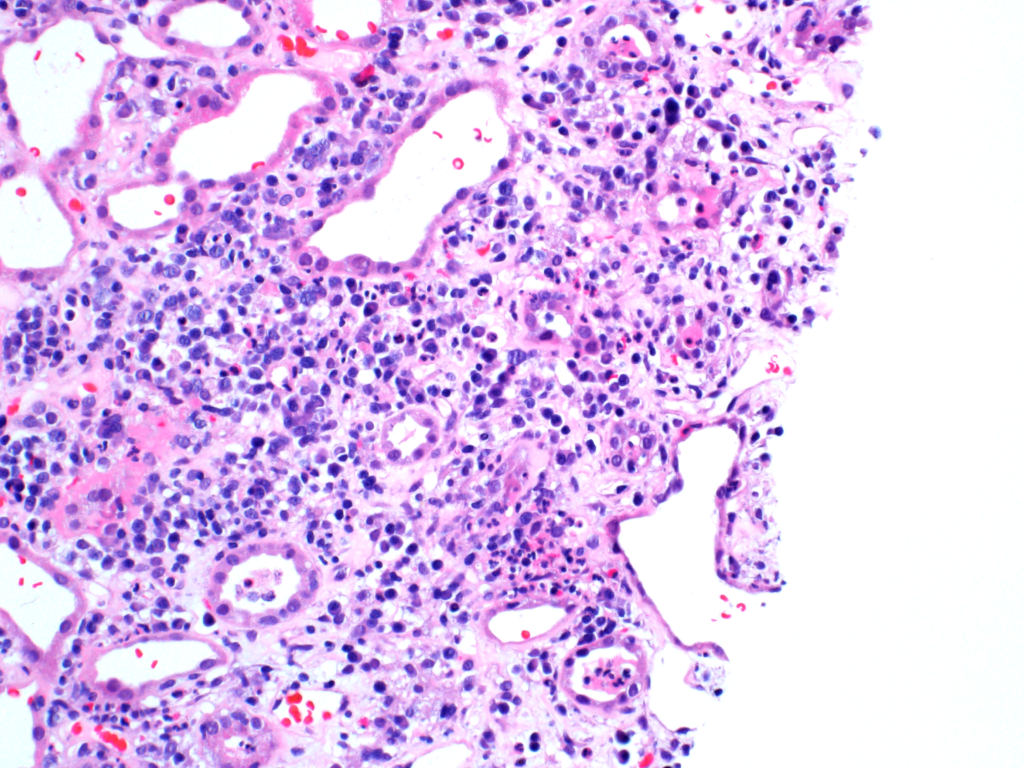
A mixed inflammatory cell infiltrate in a child with acute interstitial nephritis. Image courtesy of Patrick Walker, MD.

Tubular Diseases acute interstitial nephritis, light microscopy

WBC casts

10 Jun 2020

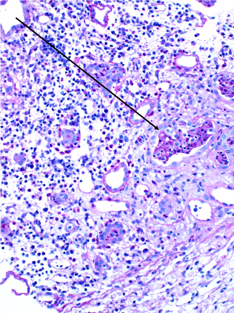
WBC casts (black arrows) in a biopsy specimen of a patient with acute interstitial nephritis. Images courtesy of Patrick Walker, MD.

General Renal Pathology, Tubular Diseases acute interstitial nephritis, light microscopy, WBC casts

Autosomal recessive polycystic kidney disease (ARPKD)

10 Jun 2020

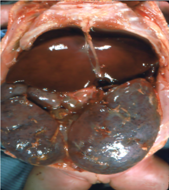
Gross pathology and low-power light microscopy of kidney tissue in a neonate with ARPKD. The kidneys are enlarged but maintain their reniform shape, and are full of microscopic cysts derived from dilated distal tubules and cortical collecting ducts. Images courtesy of Patrick Walker, MD.


Genetic Diseases, Tubular Diseases gross pathology, light microscopy, polycystic kidney disease, renal cysts

Simple renal cysts

10 Jun 2020

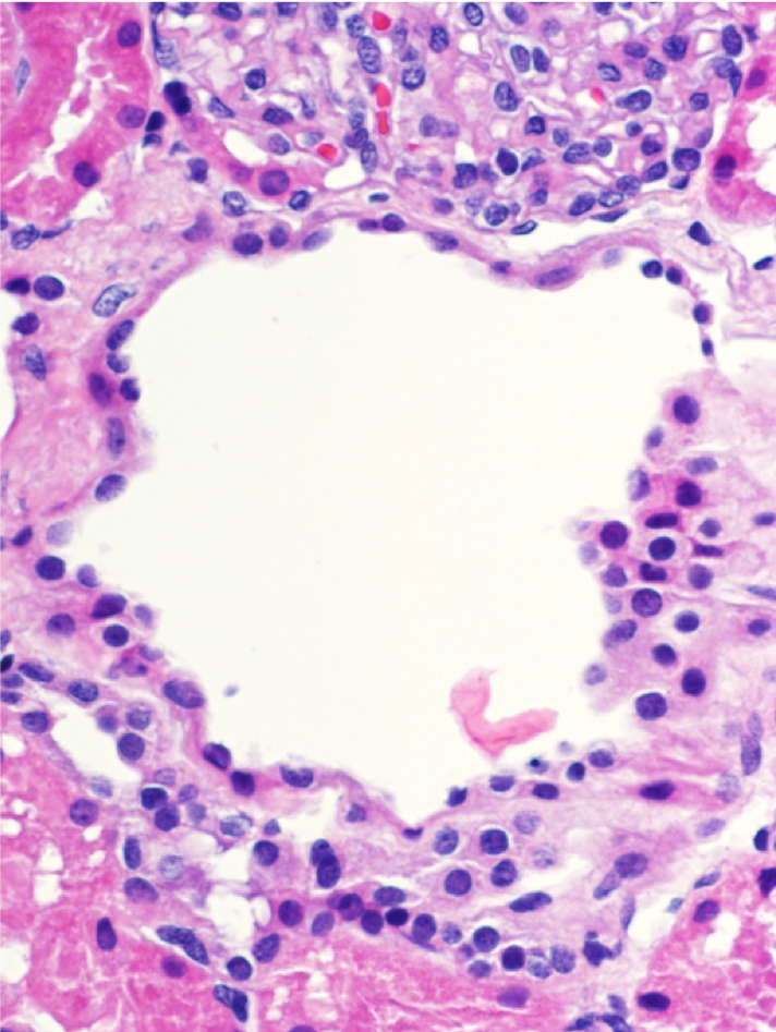
Microscopic renal cysts. Note the flattened to cuboidal epithelium lining the cysts. Images courtesy of Patrick Walker, MD.

General Renal Pathology, Tubular Diseases light microscopy, renal cysts

Search the Site

6728 Old McLean Village Dr., McLean, VA 22101 Tel 703.556.9222 fax 703.556.8729 © ASPN 2018