Archive for 2020
Simple renal cysts
Microscopic renal cysts. Note the flattened to cuboidal epithelium lining the cysts. Images courtesy of Patrick Walker, MD.
Read MoreCrescents
A segmental (left, black arrow) and circumferential crescent (right) in a patient with IgA nephropathy. Images courtesy of Patrick Walker, MD.
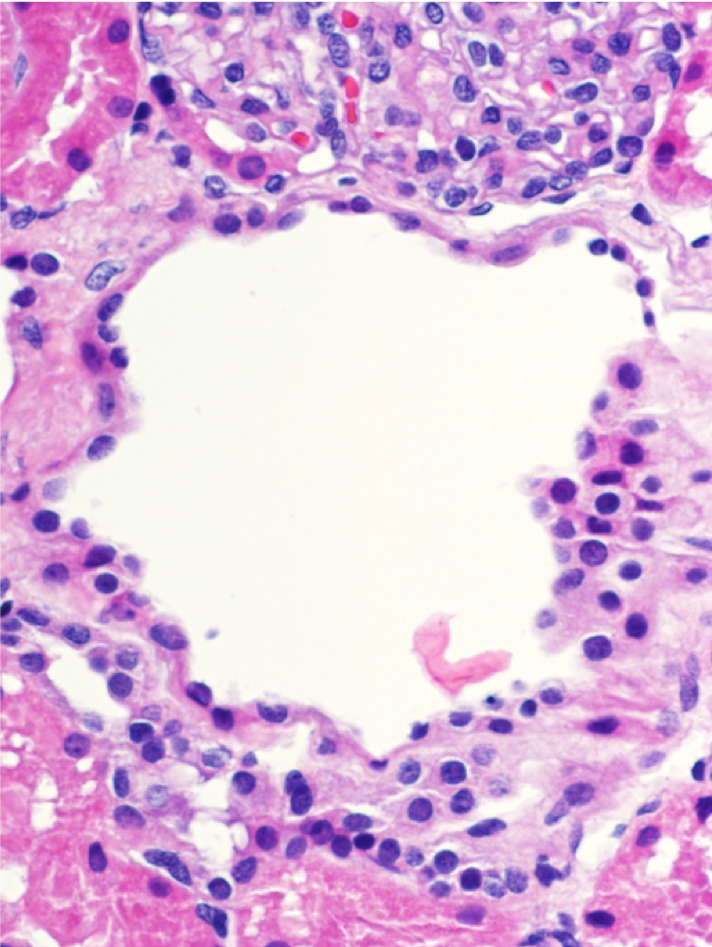
Read MoreEndocapillary hypercellularity
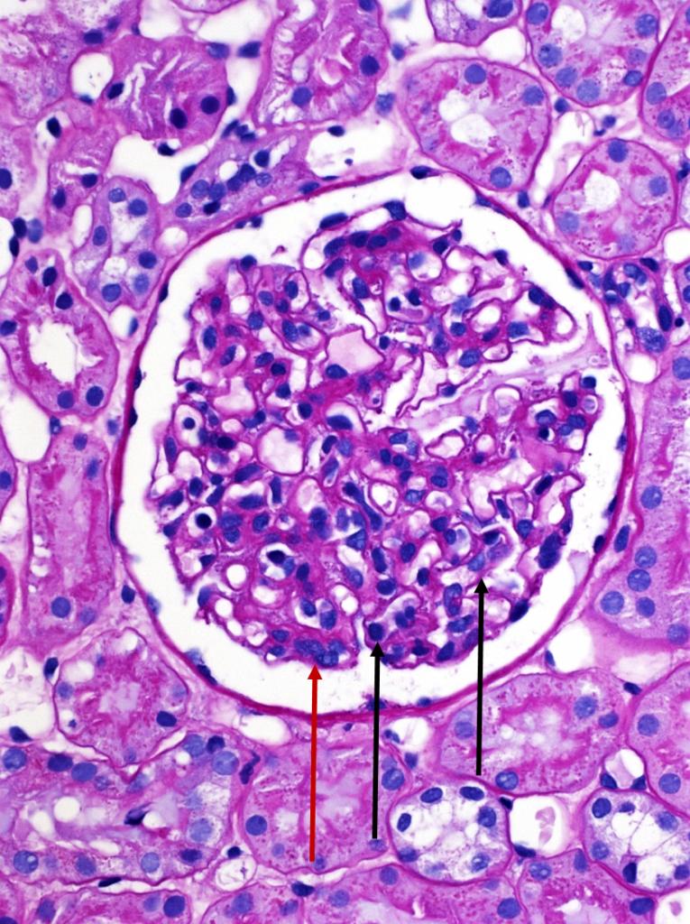
A normal appearing glomerulus (left) compared to a glomerulus with endocapillary hypercellularity (right). Note the hypercellular capillary loop (red arrow) compared to the normal capillary lumens (black arrows). This histologic…
Read MoreImmunofluorescent staining in C3 glomerulopathy
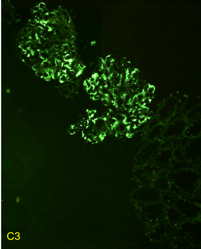
Immunofluorescent staining pattern in a patient with C3 glomerulopathy. There is strong C3 staining in the capillary loops and mesangium. Immunoglobulin staining, such as IgG, is typically absent or at…
Read MoreDense deposit disease
Dense deposit disease on electron microscopy in a patient with C3 glomerulopathy. This lesion results from intramembranous transformation of the glomerular basement membrane by sausage-like, “osmiophilic” dense material. Images courtesy…
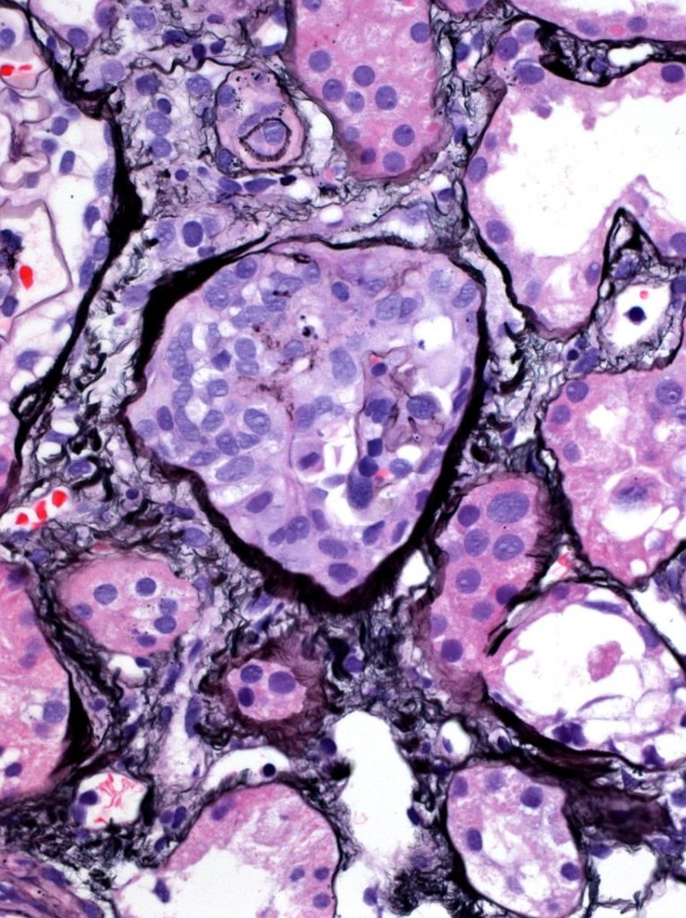
Read MoreMembranoproliferative glomerulonephritis

Membranoproliferative pattern of glomerular injury in a patient with C3 glomerulopathy. This pattern of injury typically has endocapillary proliferation, diffuse capillary wall thickening, increased mesangial matrix, and mesangial proliferation visible…
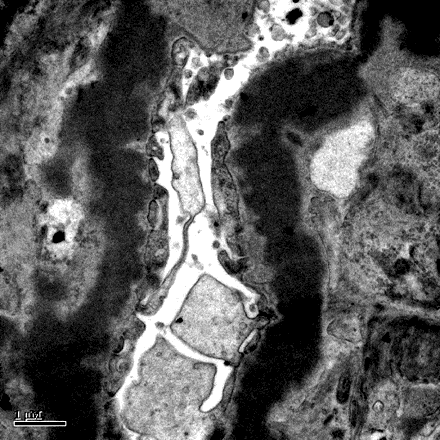
Read MoreFoam Cells
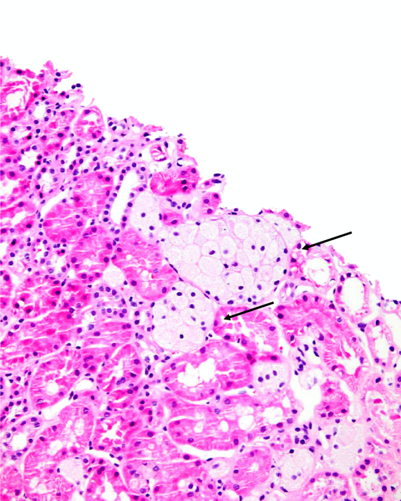
Clusters of interstitial foam cells (arrows) in a kidney biopsy. These are commonly found in biopsy specimens of patients with Alport syndrome, FSGS, IgA nephropathy, and other proteinuric kidney diseases.…
Read MoreASPN Statement Against Racism (June 02, 2020)
For many in our community, the events of the last week have caused great consternation and triggered feelings of intense disappointment, frustration, and anger. For others, what has transpired has…
Read MoreDivision Director’s Meeting
Duplex collecting system with 2 ureters on one side on VCUG
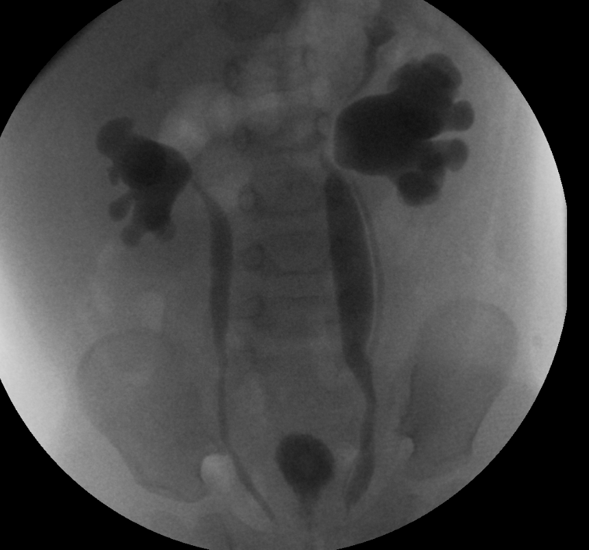
This voiding cystourethrogram shows bilateral vesicoureteral reflux grade 5. Also, on left side, two separate ureters can be seen.
Read More