Posts by Brian Stotter
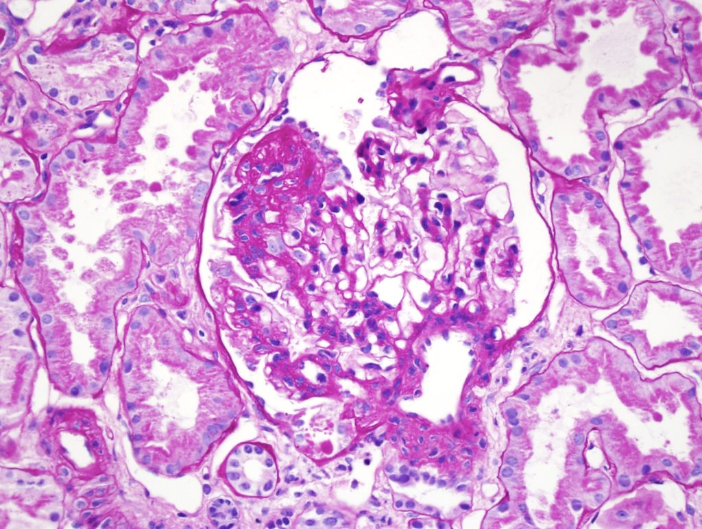
Endocapillary hypercellularity
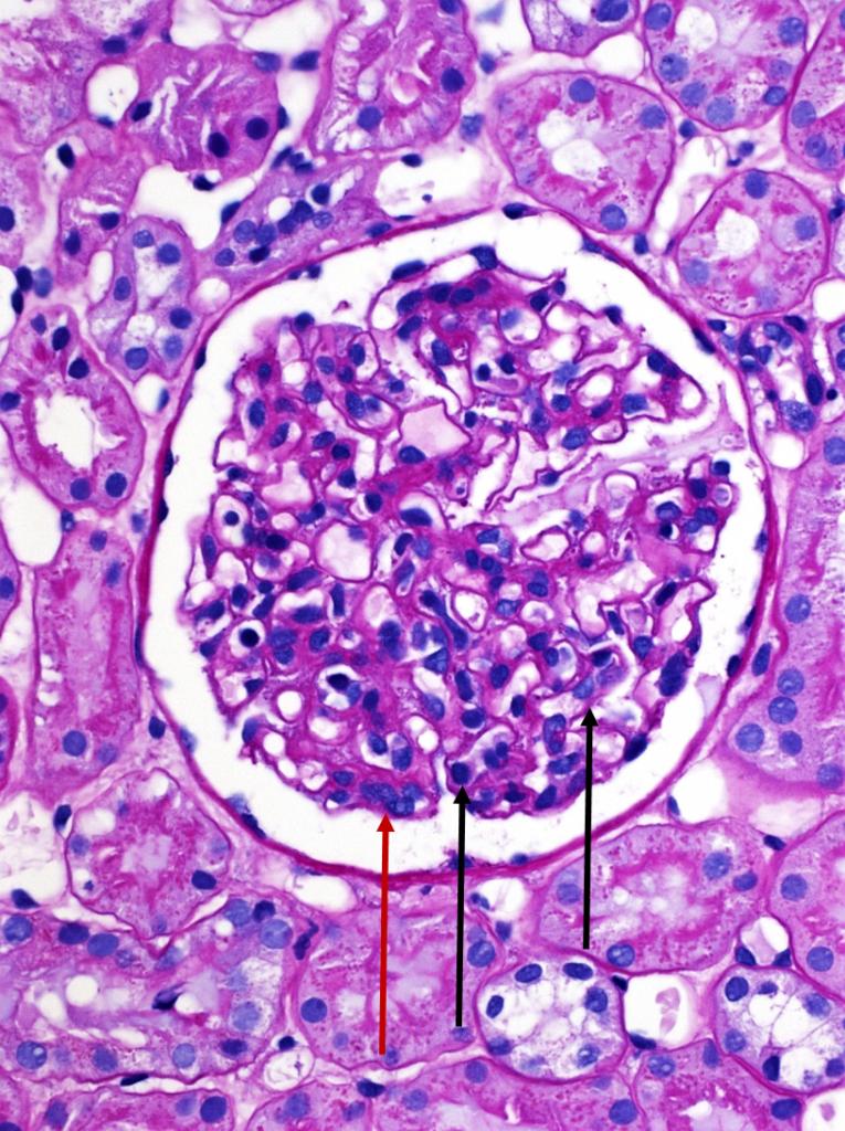
A normal appearing glomerulus (left) compared to a glomerulus with endocapillary hypercellularity (right). Note the hypercellular capillary loop (red arrow) compared to the normal capillary lumens (black arrows). This histologic…
Read MoreImmunofluorescent staining in C3 glomerulopathy
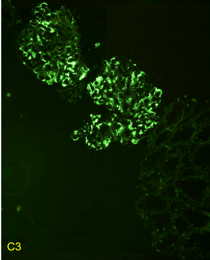
Immunofluorescent staining pattern in a patient with C3 glomerulopathy. There is strong C3 staining in the capillary loops and mesangium. Immunoglobulin staining, such as IgG, is typically absent or at…
Read MoreDense deposit disease
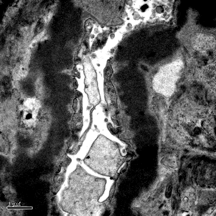
Dense deposit disease on electron microscopy in a patient with C3 glomerulopathy. This lesion results from intramembranous transformation of the glomerular basement membrane by sausage-like, “osmiophilic” dense material. Images courtesy…
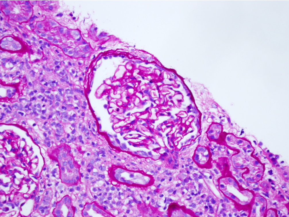
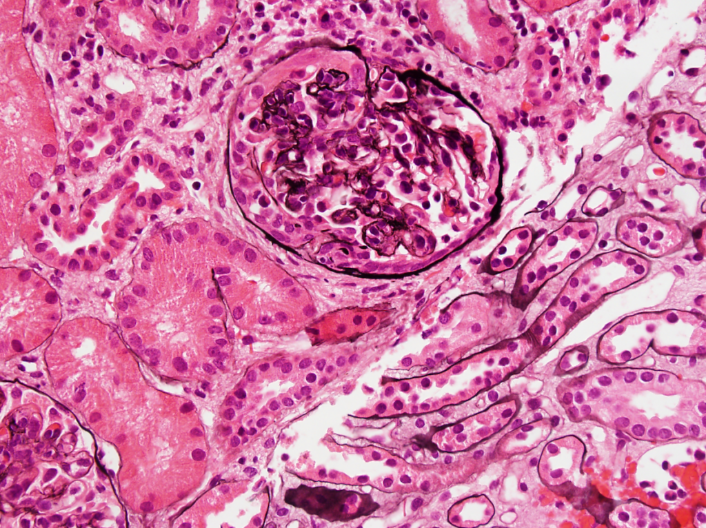
Read MoreMembranoproliferative glomerulonephritis

Membranoproliferative pattern of glomerular injury in a patient with C3 glomerulopathy. This pattern of injury typically has endocapillary proliferation, diffuse capillary wall thickening, increased mesangial matrix, and mesangial proliferation visible…
Read MoreFoam Cells
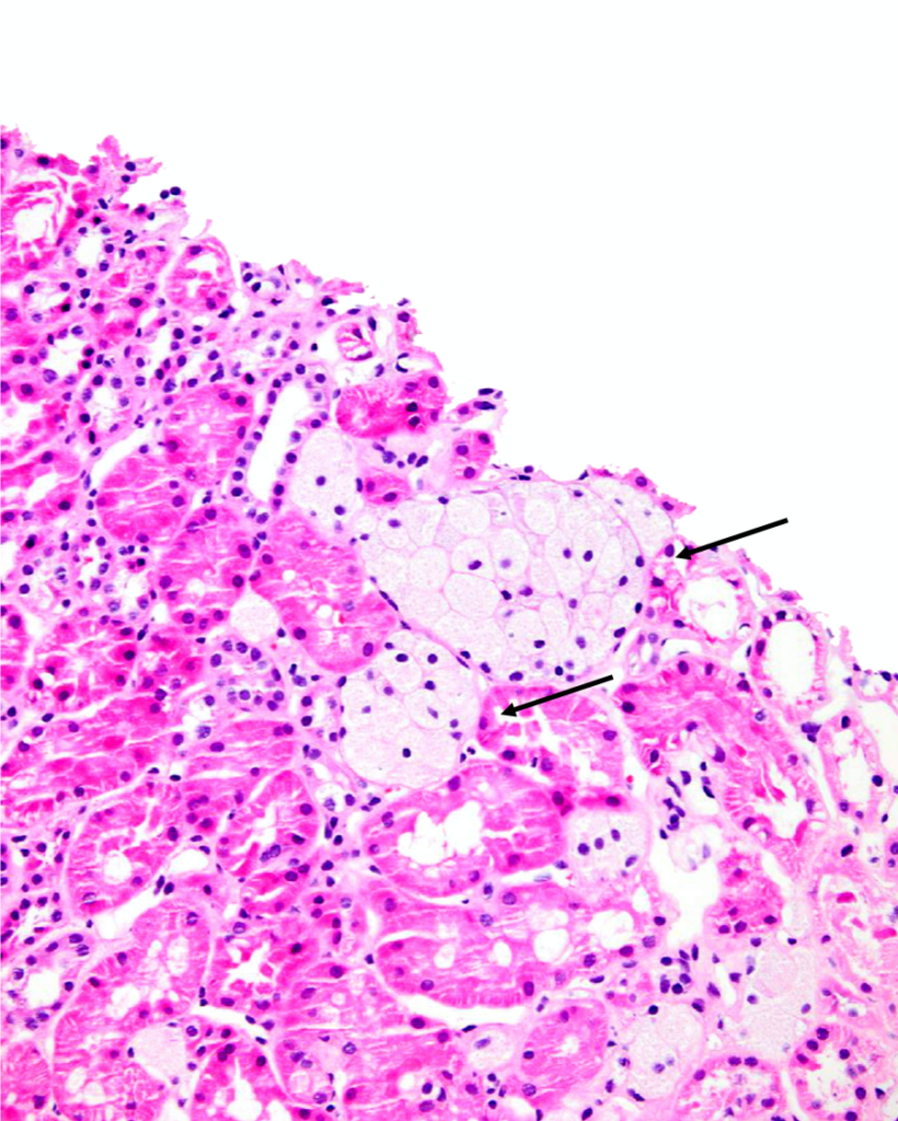
Clusters of interstitial foam cells (arrows) in a kidney biopsy. These are commonly found in biopsy specimens of patients with Alport syndrome, FSGS, IgA nephropathy, and other proteinuric kidney diseases.…
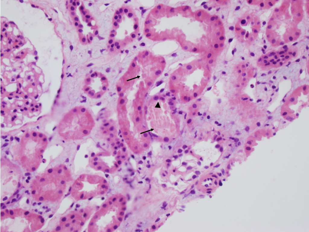
Read MoreAcute interstitial nephritis
Acute interstitial nephritis with associated acute tubular injury. There is interstitial edema and the tubules are not back to back as would be expected, due to the inflammatory and lymphocytic…
Read MoreFocal segmental glomerulosclerosis
Focal segmental glomerulosclerosis (FSGS) in a child presenting with nephrotic syndrome. Image courtesy of Joseph Gaut, MD PhD.
Read MoreMinimal change disease
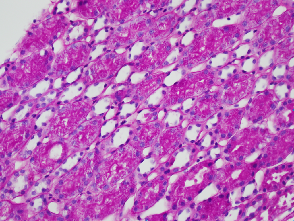
Numerous PAS-positive protein reabsorption droplets in the renal tubules of a child with minimal change disease. Image courtesy of Joseph Gaut, MD PhD.
Read MoreAcute tubular necrosis
Acute tubular necrosis (ATN). Note the tubules are not back-to-back due to interstitial edema (Masson trichrome staining, not shown, did not show appreciable fibrosis). There is blebbing and sloughing of…
Read MoreIgA nephropathy
IgA nephropathy with crescents. Note the fibrocellular crescent present in this glomerulus extending from the 3 o’clock the 12 o’clock position. Though difficult to appreciate in this image, there is…
Read More