Posts by Vikas Dharnidharka
Nuclear medicine imaging scan from back, showing a left pelvic kidney
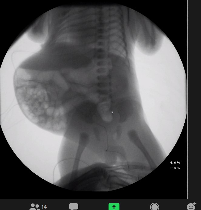
KUB showing Prune Belly abdomen
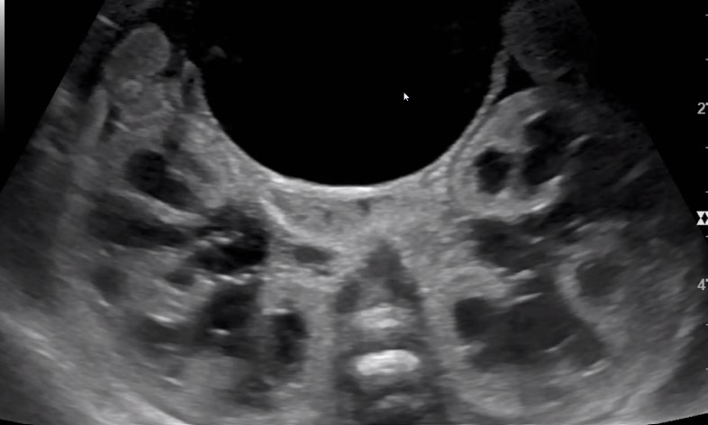
Prenatal ultrasound showing distended bilateral renal hydronephrosis and distended urinary bladder (12 o’clock)
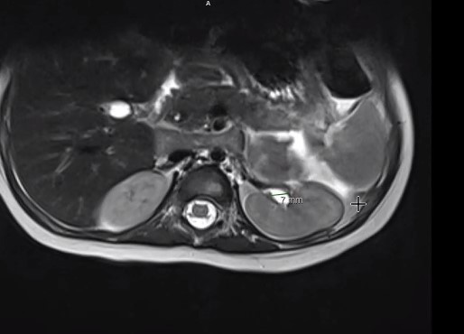
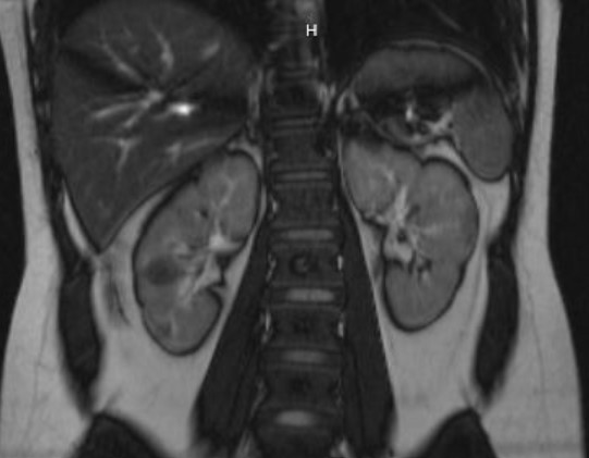
MRI image left kidney proximal ureter dilatation
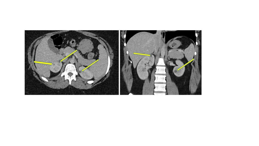
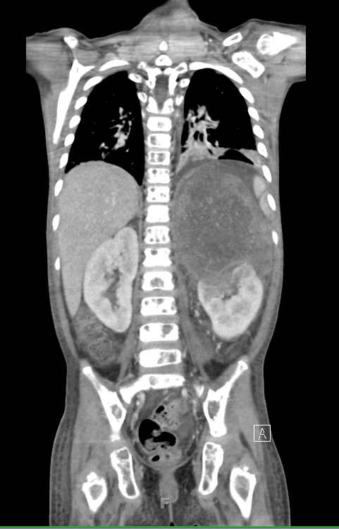
CT scan showing bilateral renal cysts (arrows) in normal sized kidneys
CT scan coronal view with left ureter 2 mm stone (arrow)
MRI coronal view angiomyolipomas or hamartomas of tuberous sclerosis in right kidney
MRI image of left kidney lower pole angiomyolipoma of tuberous sclerosis

Duplex collecting system with 2 ureters on one side on VCUG
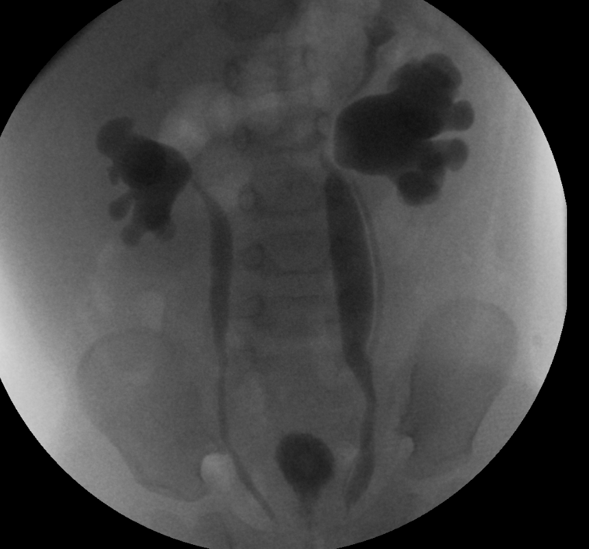
This voiding cystourethrogram shows bilateral vesicoureteral reflux grade 5. Also, on left side, two separate ureters can be seen.
Read MoreLarge neuroblastoma above left kidney CT scan coronal view
Large neuroblastoma mass above left kidney, distorting its architecture
Read More