General Renal Pathology
Mesangial Deposits
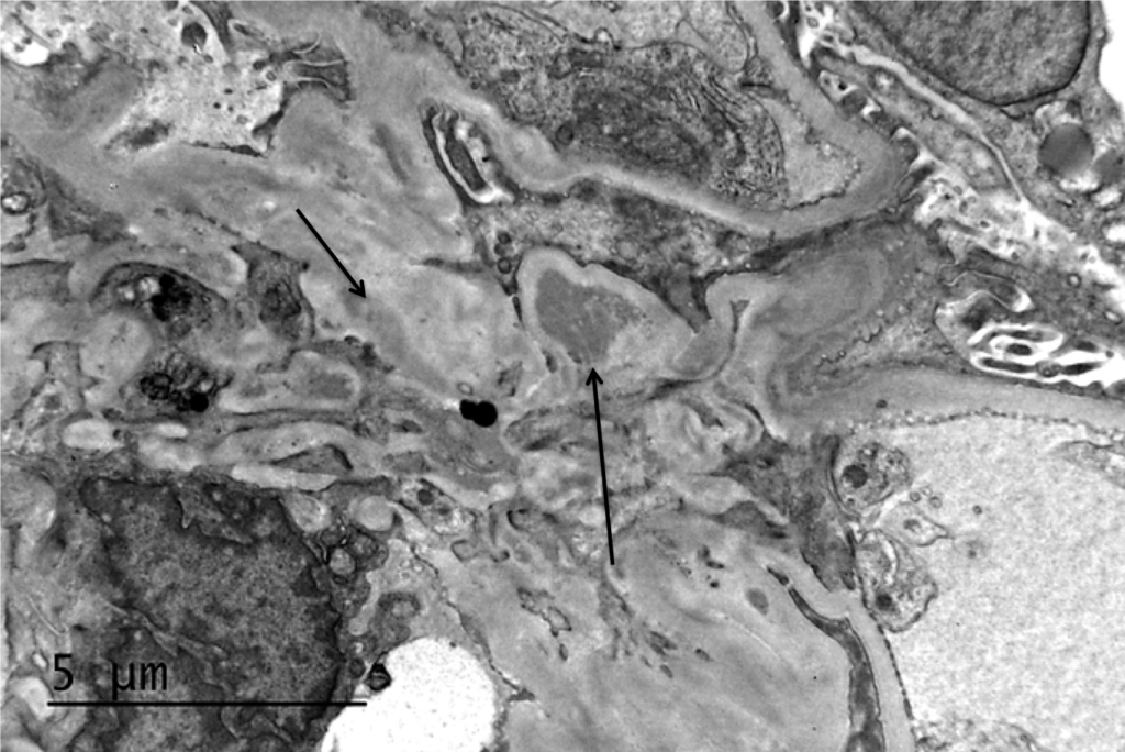
Electron microscopy of a biopsy specimen in a patient with IgA nephropathy. Electron dense deposits can be identified in the mesangium (black arrows), which on immunofluorescence would have predominant or…
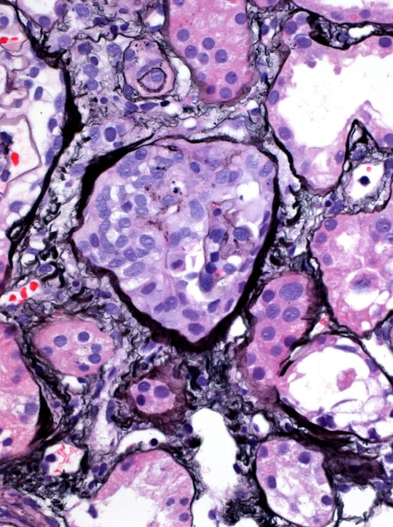
Read MoreGlomerulomegaly
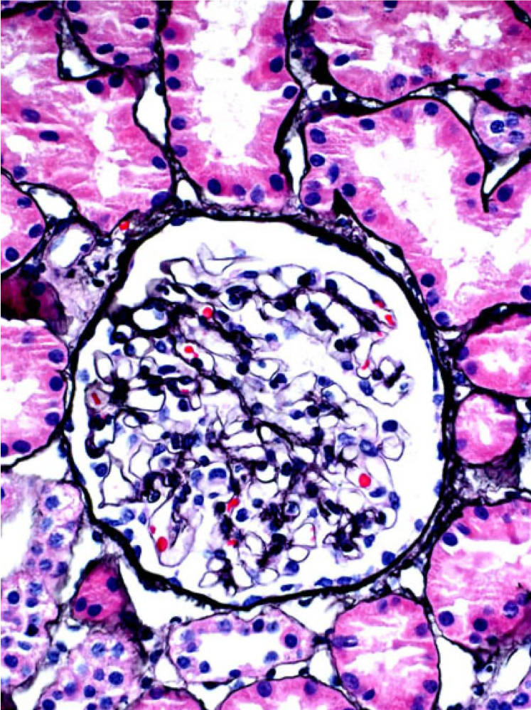
A normal glomerulus (left) and hypertrophied glomerulus (glomerulomegaly, right). Glomerulomegaly is an adaptive response to decreased nephron number (e.g. prematurity) and/or increased demand (e.g. obesity). Patients with glomerulomegaly may have…
Read MoreWBC casts
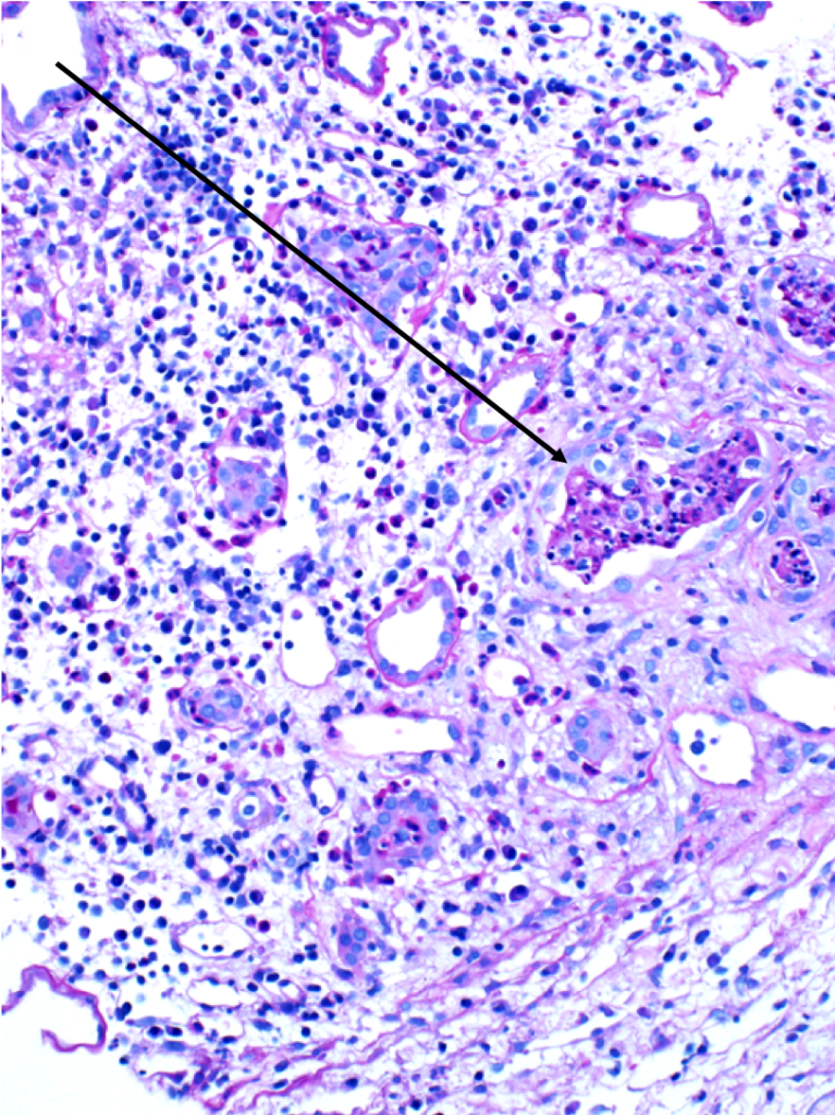
WBC casts (black arrows) in a biopsy specimen of a patient with acute interstitial nephritis. Images courtesy of Patrick Walker, MD.
Read MoreSimple renal cysts
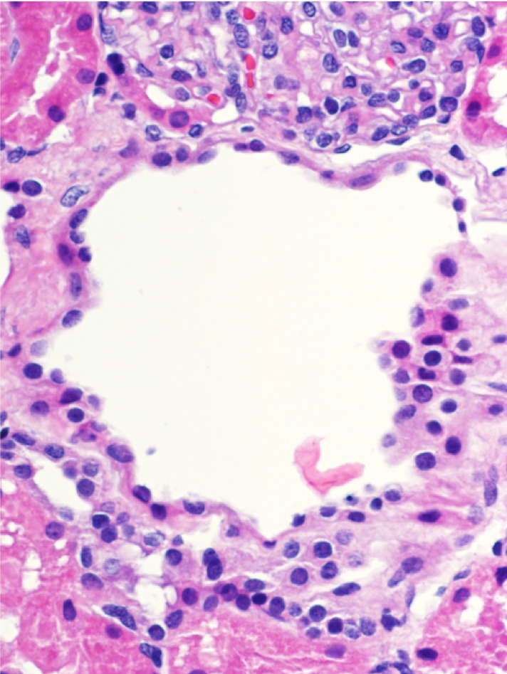
Microscopic renal cysts. Note the flattened to cuboidal epithelium lining the cysts. Images courtesy of Patrick Walker, MD.
Read MoreCrescents
A segmental (left, black arrow) and circumferential crescent (right) in a patient with IgA nephropathy. Images courtesy of Patrick Walker, MD.
Read MoreEndocapillary hypercellularity
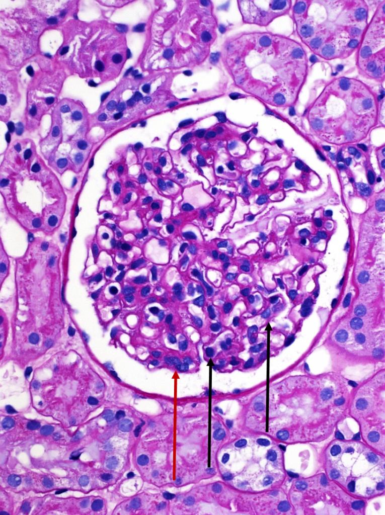
A normal appearing glomerulus (left) compared to a glomerulus with endocapillary hypercellularity (right). Note the hypercellular capillary loop (red arrow) compared to the normal capillary lumens (black arrows). This histologic…
Read MoreFoam Cells
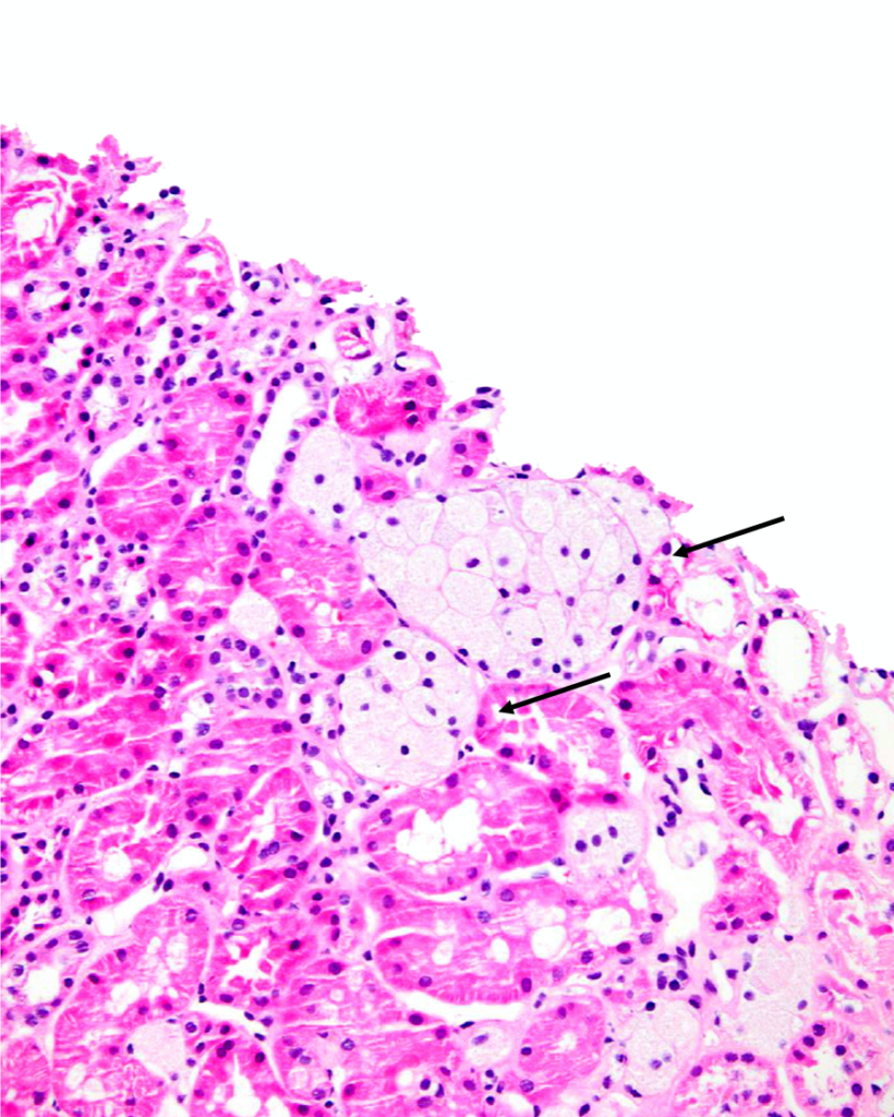
Clusters of interstitial foam cells (arrows) in a kidney biopsy. These are commonly found in biopsy specimens of patients with Alport syndrome, FSGS, IgA nephropathy, and other proteinuric kidney diseases.…
Read More