Glomerular Diseases
Minimal change disease
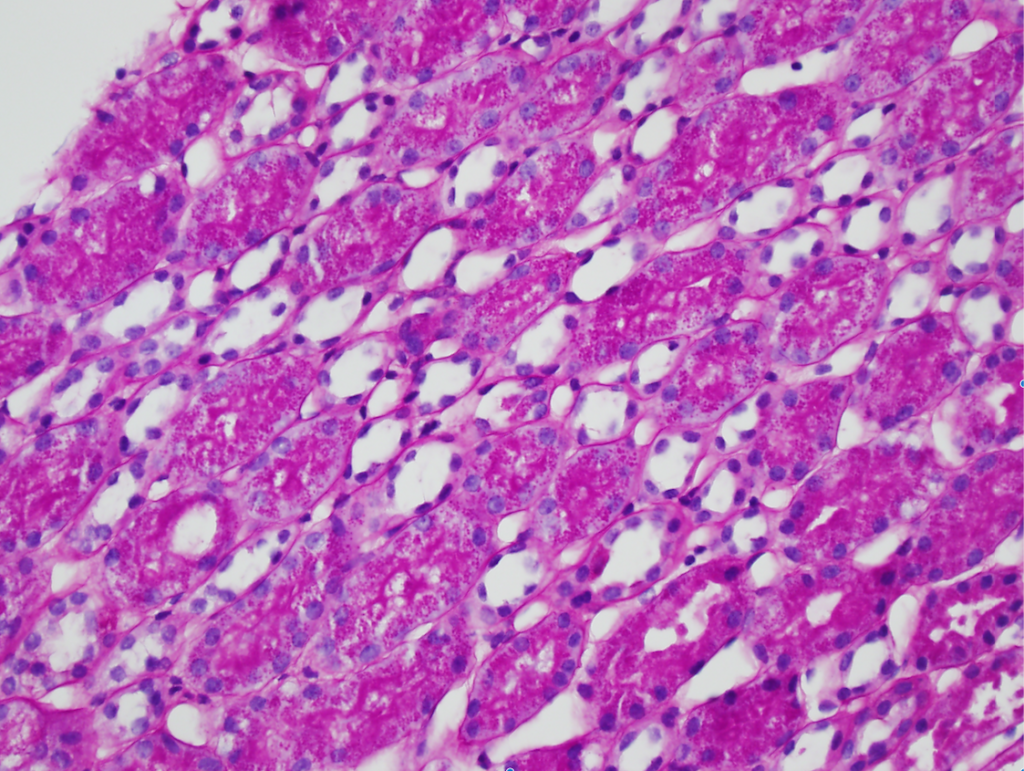
Numerous PAS-positive protein reabsorption droplets in the renal tubules of a child with minimal change disease. Image courtesy of Joseph Gaut, MD PhD.
Read MoreIgA nephropathy
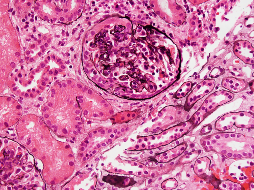
IgA nephropathy with crescents. Note the fibrocellular crescent present in this glomerulus extending from the 3 o’clock the 12 o’clock position. Though difficult to appreciate in this image, there is…
Read MoreSLE nephritis
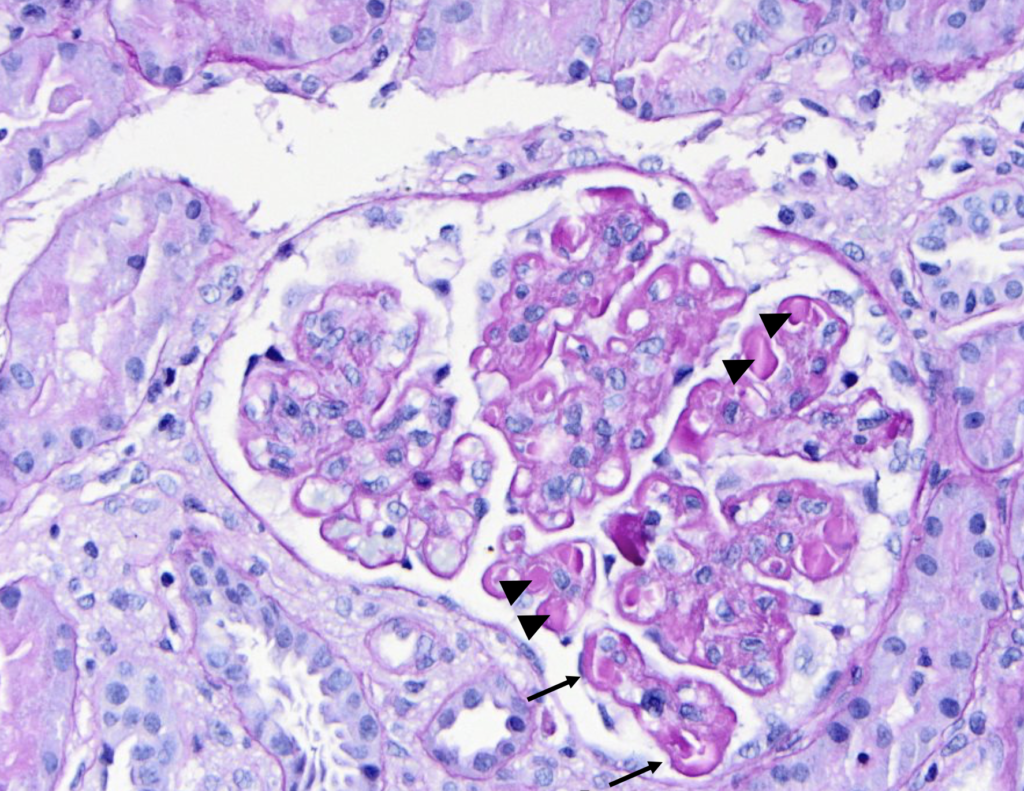
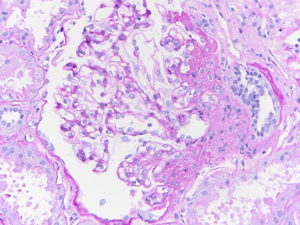
PAS stain of a glomerulus in a patient with SLE nephritis. There is endocapillary and mesangial proliferation, as evidenced by thickened, occlusive capillary loops and increased mesangial cellularity. Note the…
Read MoreTubuloreticular inclusions in SLE nephritis
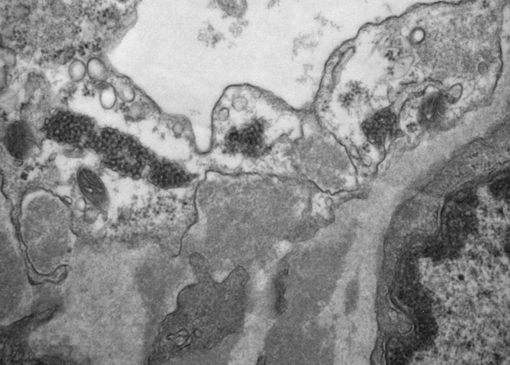
Tubuloreticular inclusions in a patient with diffuse proliferative SLE nephritis (SLE class IV). These subcellular structures (dark circular clusters) on transmission electron microscopy are localized to the cytoplasm of endothelial…
Read MoreFocal and segmental glomerulosclerosis
Segmental obliteration of the glomerular capillary lumen in a patient with FSGS. Note the sclerotic portion of the glomerular tuft is adherent to Bowman’s capsule. There is proximal tubular hypertrophy,…
Read More