Other radiology
Nuclear medicine imaging scan from back, showing a left pelvic kidney
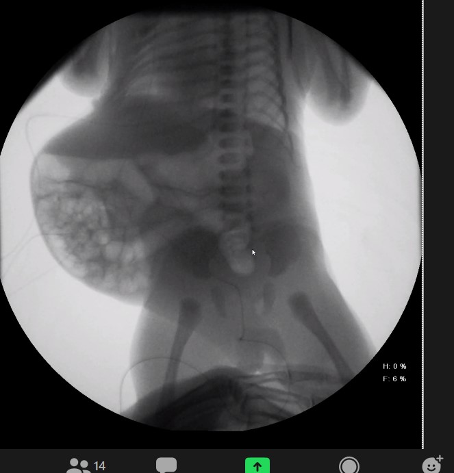
KUB showing Prune Belly abdomen
Rickets
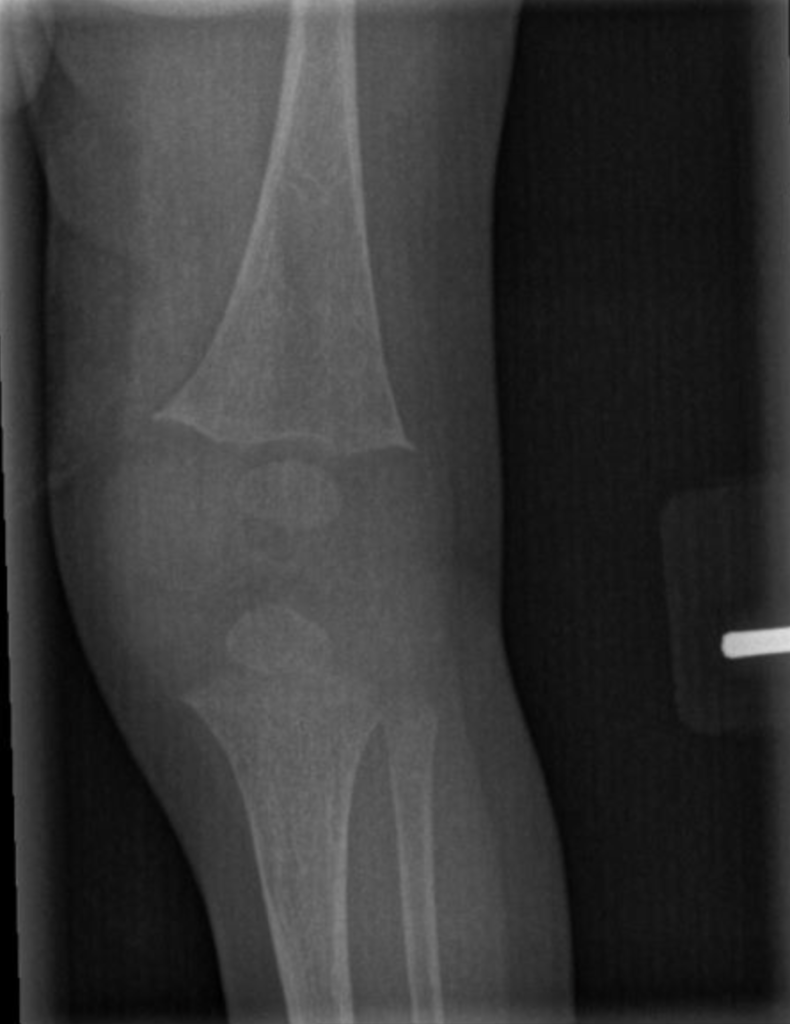
Early radiographic changes on left knee X-ray in an infant with hypophosphatemic rickets. There is decreased mineralization of the long bones and splaying, or widening, at the distal femur and…
Read More