Radiology Image Bank
| Image | Title | Summary | Categories | Tags | hf:categories | hf:tags |
|---|---|---|---|---|---|---|
| Nuclear medicine imaging scan from back, showing a left pelvic kidney | … | Other radiology | other-radiology | |||
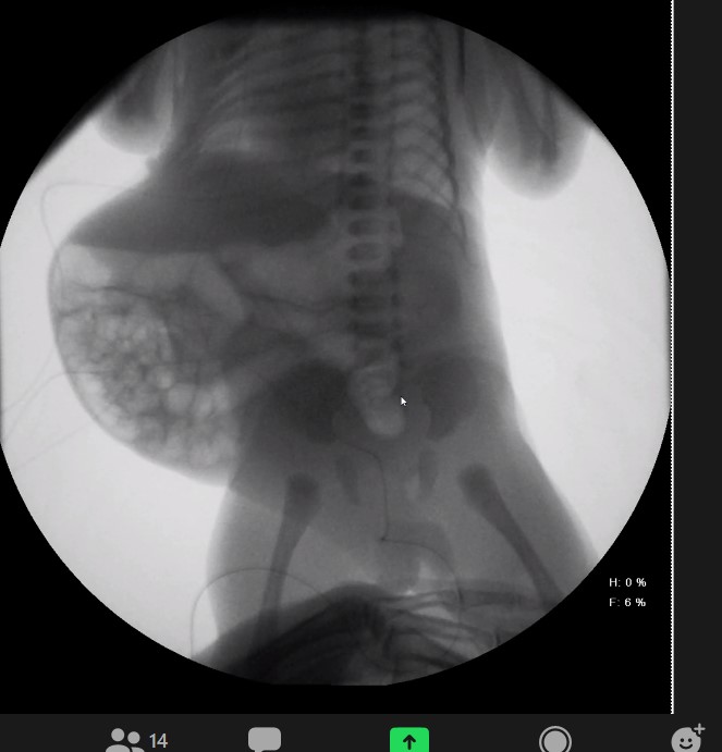
| KUB showing Prune Belly abdomen | … | Other radiology | other-radiology | |||
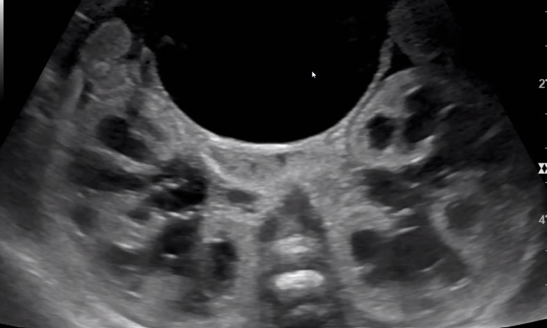
| Prenatal ultrasound showing distended bilateral renal hydronephrosis and distended urinary bladder (12 o’clock) | … | Ultrasound | ultrasound | |||
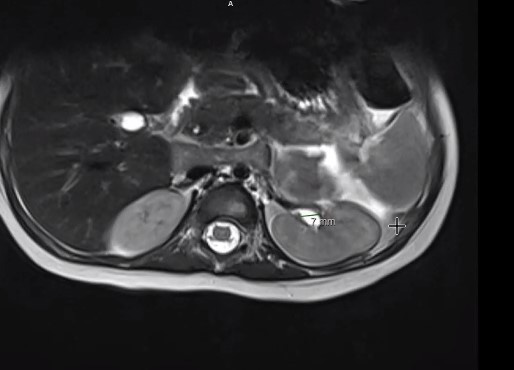
| MRI image left kidney proximal ureter dilatation | … | MRI/MRU | mri | |||
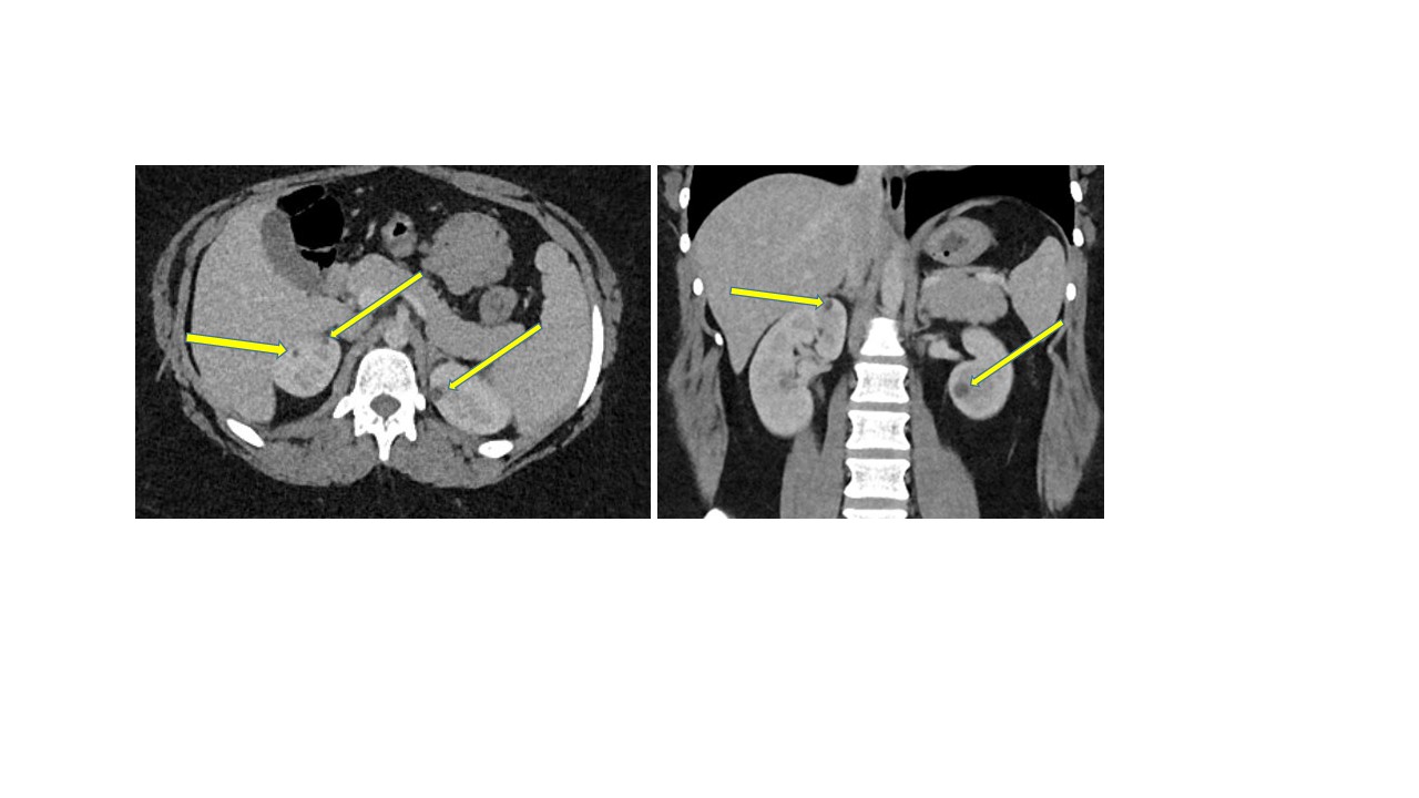
| CT scan showing bilateral renal cysts (arrows) in normal sized kidneys | … | CT | ct-scan | |||
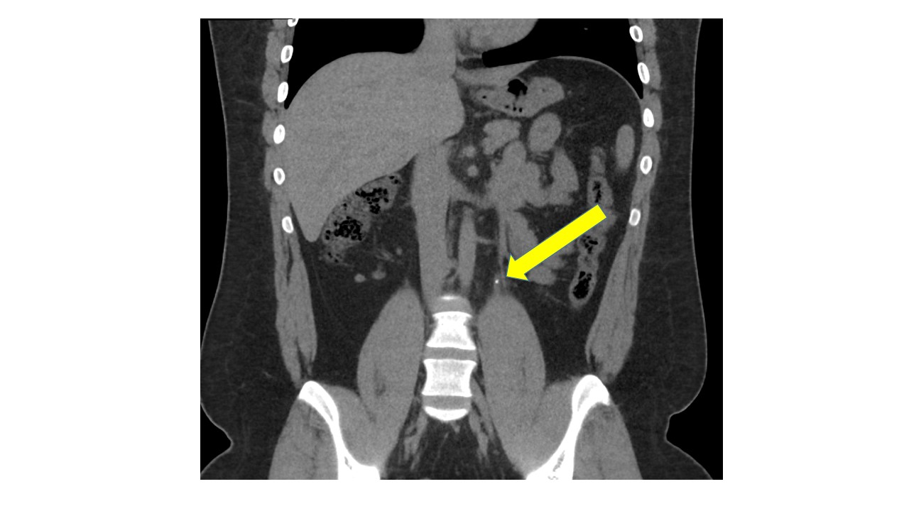
| CT scan coronal view with left ureter 2 mm stone (arrow) | … | CT | ct-scan | |||
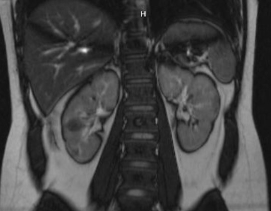
| MRI coronal view angiomyolipomas or hamartomas of tuberous sclerosis in right kidney | … | MRI/MRU | mri | |||
| MRI image of left kidney lower pole angiomyolipoma of tuberous sclerosis | … | MRI/MRU | mri | |||
| Calyceal Diverticulum on MRU | Right anterior calyceal diverticulum on functional MR Urography. Top left side shows a T2 weighted image (no contrast) where fluid is bright, top right image is 10 min post-contrast on axial plane, and bottom images are a coronal views showing filling of diverticulum on dynamic phase. Images courtesy of Hansel Otero, … | MRI/MRU | mri | |||
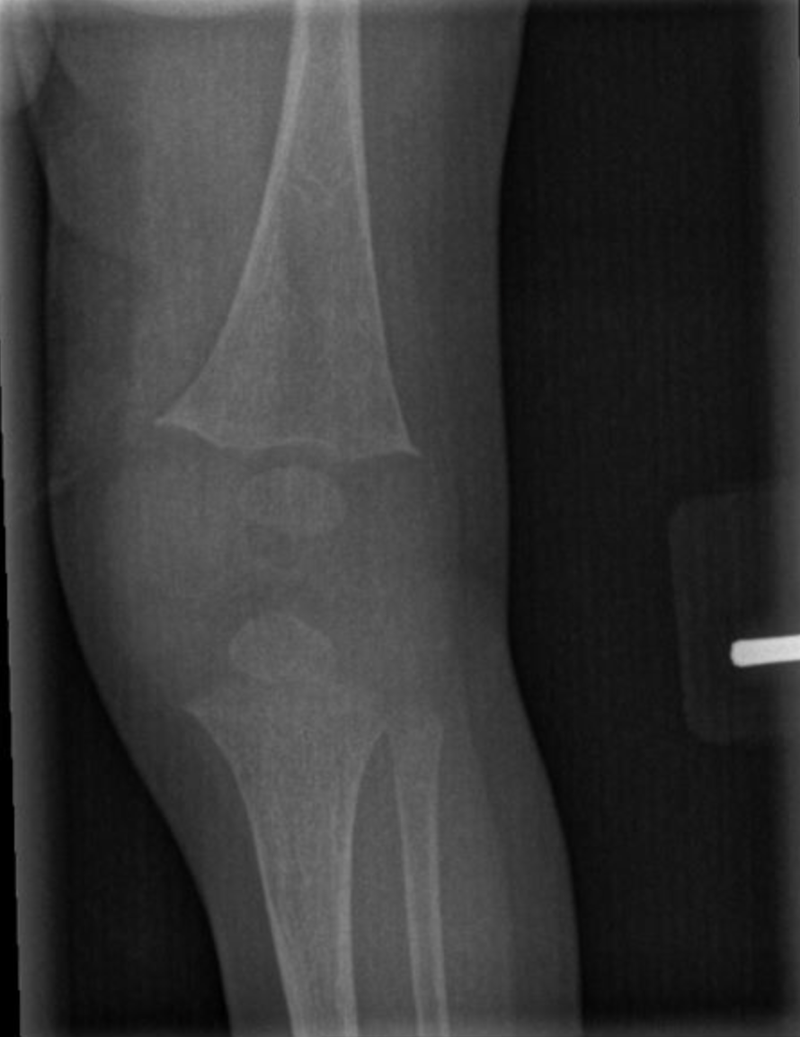
| Rickets | Early radiographic changes on left knee X-ray in an infant with hypophosphatemic rickets. There is decreased mineralization of the long bones and splaying, or widening, at the distal femur and proximal tibia, where the metaphyses are … | Other radiology | rickets, X-ray | other-radiology | rickets x-ray | |
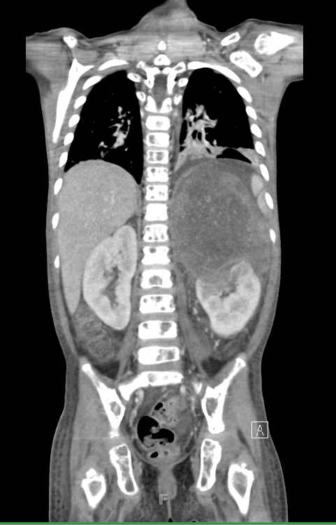
| Large neuroblastoma above left kidney CT scan coronal view | Large neuroblastoma mass above left kidney, distorting its … | CT | coronal, CT scan, left kidney, neuroblastoma | ct-scan | coronal ct-scan left-kidney neuroblastoma | |
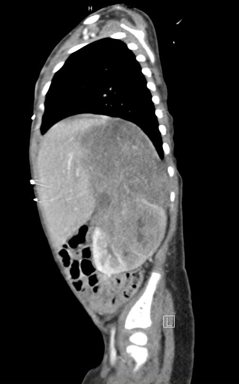
| Left kidney neuroblastoma sagittal image | This image shows a large neuroblastoma mass at upper pole of left kidney, distorting its … | CT | CT scan sagittal, left kidney, neuroblastoma | ct-scan | ct-scan-sagittal left-kidney neuroblastoma | |
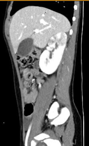
| Acute pyelonephritis on CT scan with contrast sagittal view | CT scan axial view with contrast showing right kidney upper pole defects, consistent with acute … | CT | CAKUT, Masses | ct-scan | cakut masses |


