Posts Tagged ‘electron microscopy’
Mesangial Deposits
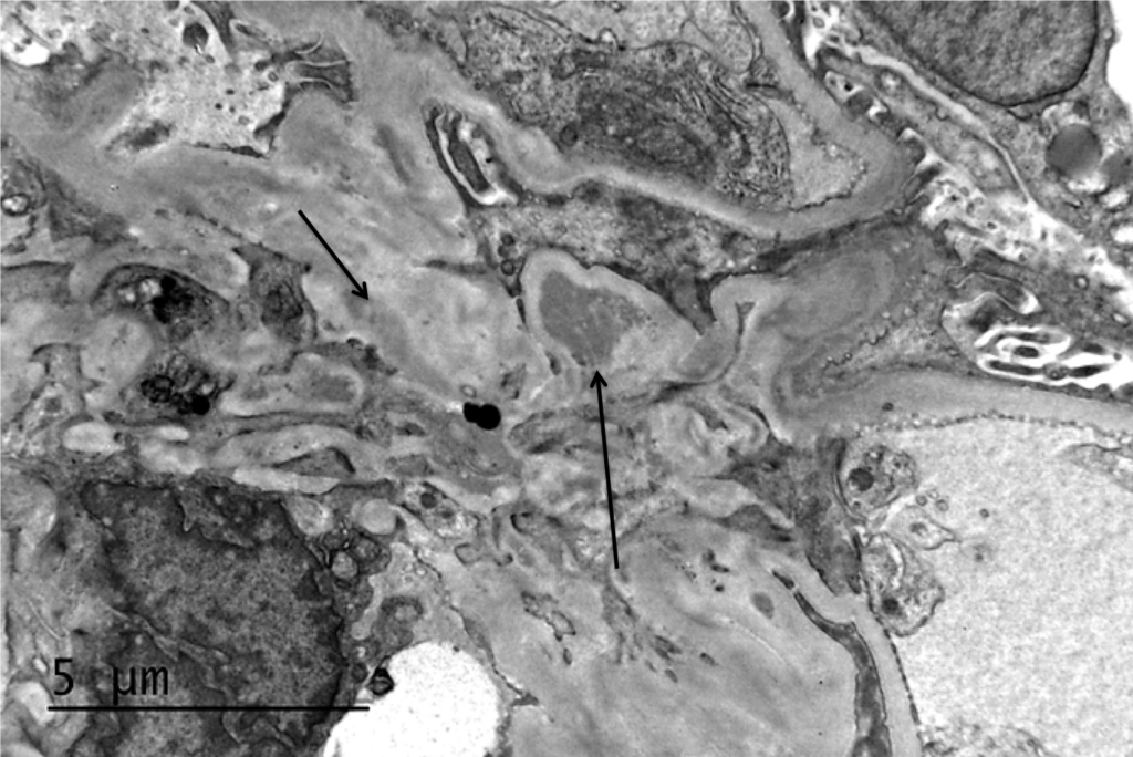
Electron microscopy of a biopsy specimen in a patient with IgA nephropathy. Electron dense deposits can be identified in the mesangium (black arrows), which on immunofluorescence would have predominant or…
Read MoreDense deposit disease
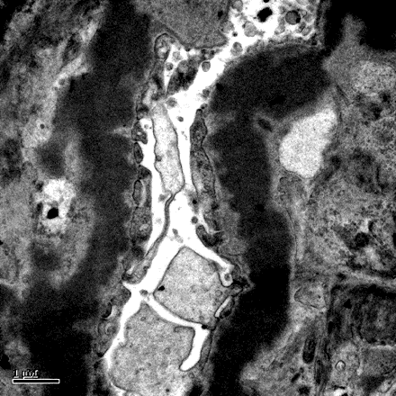
Dense deposit disease on electron microscopy in a patient with C3 glomerulopathy. This lesion results from intramembranous transformation of the glomerular basement membrane by sausage-like, “osmiophilic” dense material. Images courtesy…
Read MoreTubuloreticular inclusions in SLE nephritis
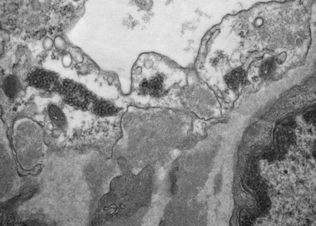
Tubuloreticular inclusions in a patient with diffuse proliferative SLE nephritis (SLE class IV). These subcellular structures (dark circular clusters) on transmission electron microscopy are localized to the cytoplasm of endothelial…
Read More