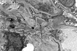
Electron microscopy of a biopsy specimen in a patient with IgA nephropathy. Electron dense deposits can be identified in the mesangium (black arrows), which on immunofluorescence would have predominant or co-dominant IgA staining. Note that although mesangial IgA deposits are classic in IgA nephropathy and IgA vasculitis (former Henoch-Schonlein purpura…
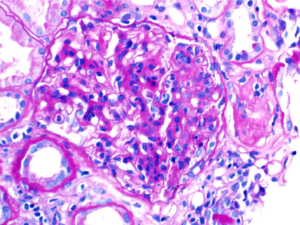
A patient with IgA nephropathy and associated segmental glomerulosclerosis. Images courtesy of Patrick Walker, MD.
A segmental (left, black arrow) and circumferential crescent (right) in a patient with IgA nephropathy. Images courtesy of Patrick Walker, MD.
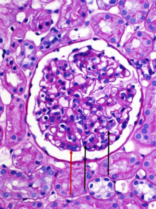
A normal appearing glomerulus (left) compared to a glomerulus with endocapillary hypercellularity (right). Note the hypercellular capillary loop (red arrow) compared to the normal capillary lumens (black arrows). This histologic feature can be seen in several glomerular disorders, including IgA nephropathy, post-infectious glomerulonephritis, lupus nephritis, and C3 glomerulopathy. Images courtesy…
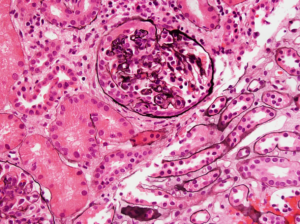
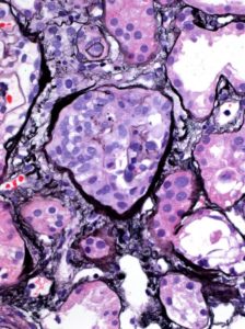
IgA nephropathy with crescents. Note the fibrocellular crescent present in this glomerulus extending from the 3 o’clock the 12 o’clock position. Though difficult to appreciate in this image, there is mild mesangial hypercellularity. Image courtesy of Joseph Gaut, MD PhD.