Posts Tagged ‘light microscopy’
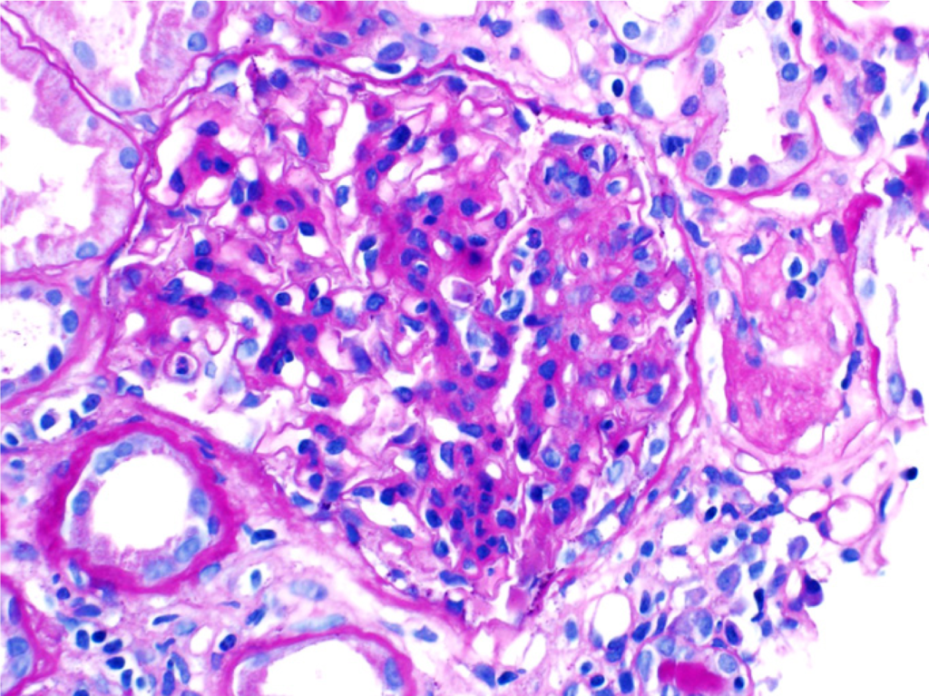
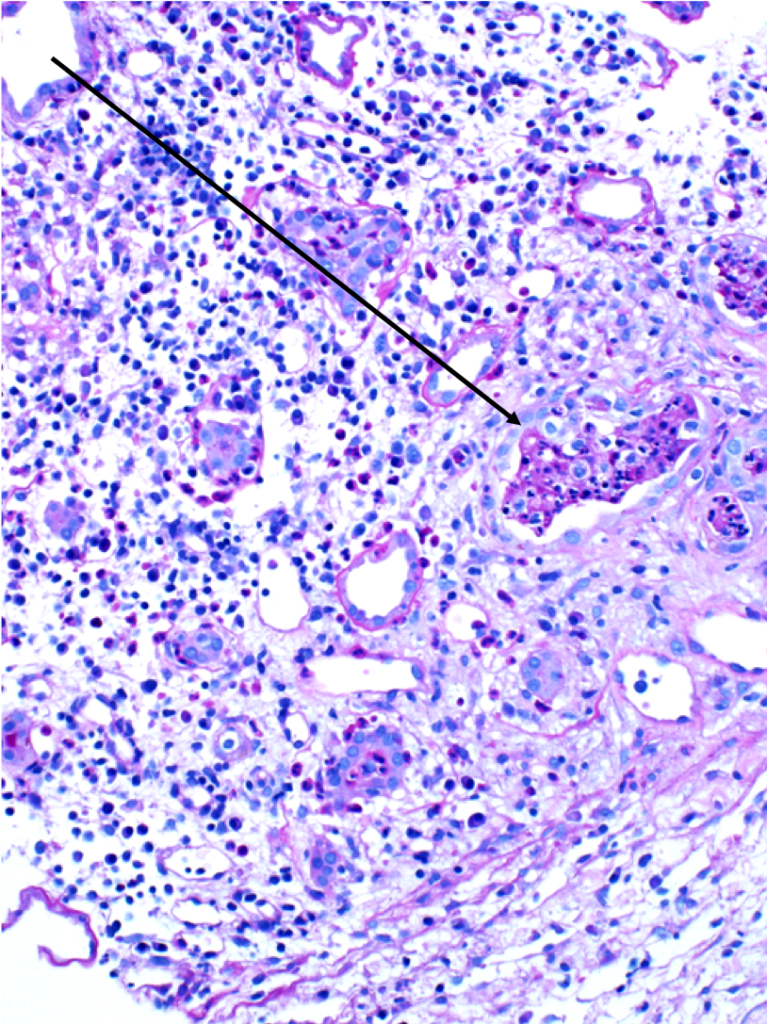
Perihilar FSGS
Perihilar variant of focal segmental glomerulosclerosis (FSGS). Hyaline deposition and sclerosis occur at the vascular pole of the glomerulus. This variant is believed to be a secondary form of FSGS,…
Read MoreGlomerulomegaly
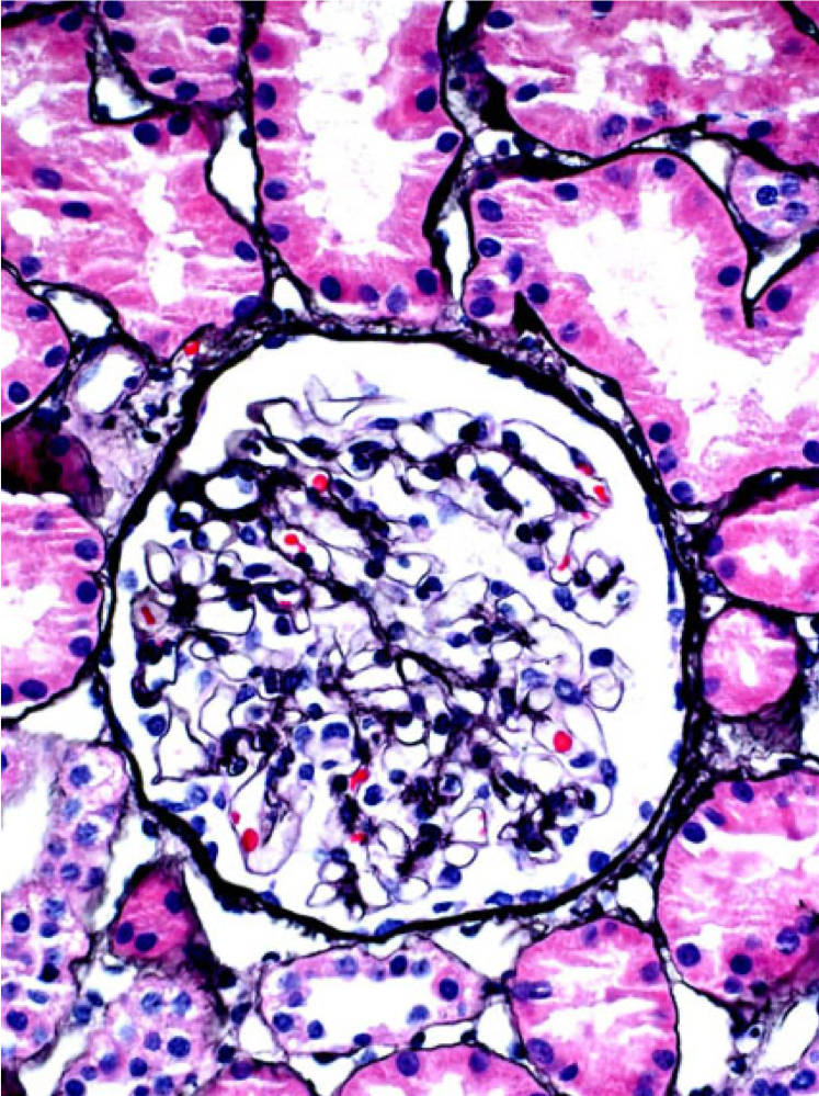
A normal glomerulus (left) and hypertrophied glomerulus (glomerulomegaly, right). Glomerulomegaly is an adaptive response to decreased nephron number (e.g. prematurity) and/or increased demand (e.g. obesity). Patients with glomerulomegaly may have…
Read MoreSegmental glomerulosclerosis in IgA nephropathy
A patient with IgA nephropathy and associated segmental glomerulosclerosis. Images courtesy of Patrick Walker, MD.
Read MoreAcute interstitial nephritis
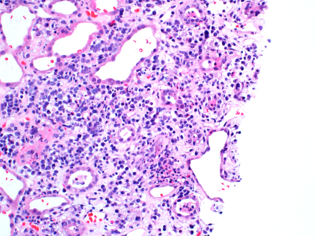
A mixed inflammatory cell infiltrate in a child with acute interstitial nephritis. Image courtesy of Patrick Walker, MD.
Read MoreWBC casts
WBC casts (black arrows) in a biopsy specimen of a patient with acute interstitial nephritis. Images courtesy of Patrick Walker, MD.
Read MoreAutosomal recessive polycystic kidney disease (ARPKD)
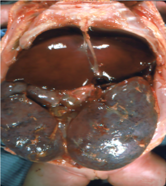
Gross pathology and low-power light microscopy of kidney tissue in a neonate with ARPKD. The kidneys are enlarged but maintain their reniform shape, and are full of microscopic cysts derived…
Read MoreSimple renal cysts
Microscopic renal cysts. Note the flattened to cuboidal epithelium lining the cysts. Images courtesy of Patrick Walker, MD.
Read MoreCrescents
A segmental (left, black arrow) and circumferential crescent (right) in a patient with IgA nephropathy. Images courtesy of Patrick Walker, MD.
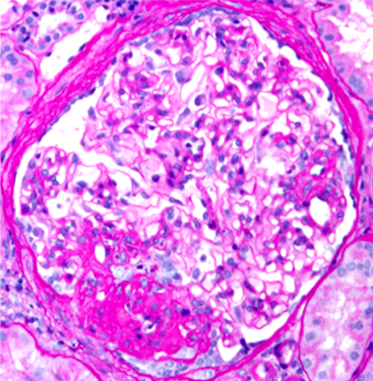
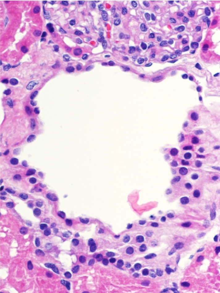
Read MoreEndocapillary hypercellularity
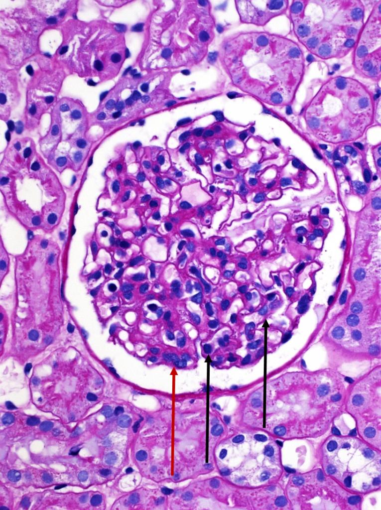
A normal appearing glomerulus (left) compared to a glomerulus with endocapillary hypercellularity (right). Note the hypercellular capillary loop (red arrow) compared to the normal capillary lumens (black arrows). This histologic…
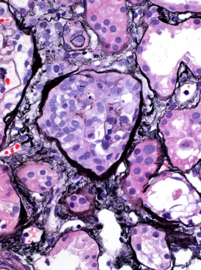
Read MoreMembranoproliferative glomerulonephritis

Membranoproliferative pattern of glomerular injury in a patient with C3 glomerulopathy. This pattern of injury typically has endocapillary proliferation, diffuse capillary wall thickening, increased mesangial matrix, and mesangial proliferation visible…
Read More