Posts Tagged ‘light microscopy’
Foam Cells
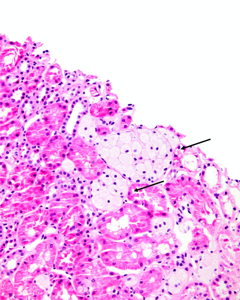
Clusters of interstitial foam cells (arrows) in a kidney biopsy. These are commonly found in biopsy specimens of patients with Alport syndrome, FSGS, IgA nephropathy, and other proteinuric kidney diseases.…
Read MoreAcute interstitial nephritis
Acute interstitial nephritis with associated acute tubular injury. There is interstitial edema and the tubules are not back to back as would be expected, due to the inflammatory and lymphocytic…
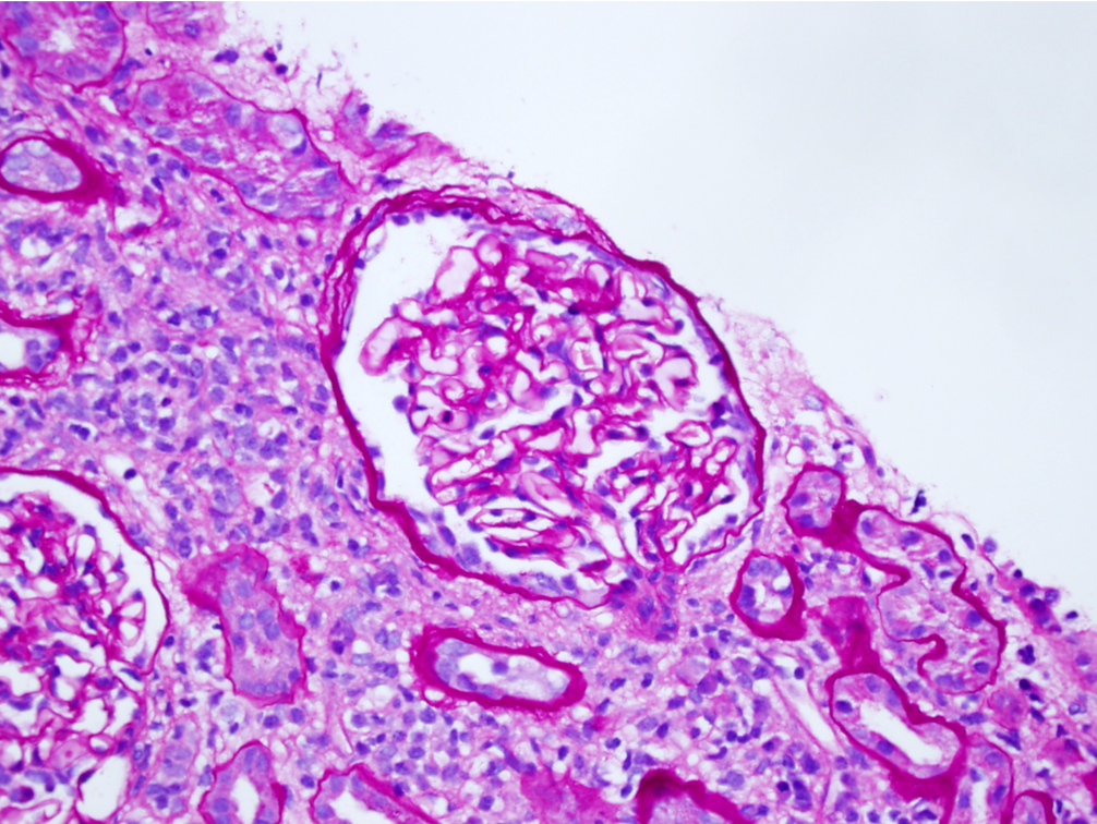
Read MoreFocal segmental glomerulosclerosis
Focal segmental glomerulosclerosis (FSGS) in a child presenting with nephrotic syndrome. Image courtesy of Joseph Gaut, MD PhD.
Read MoreMinimal change disease
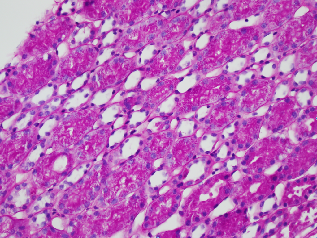
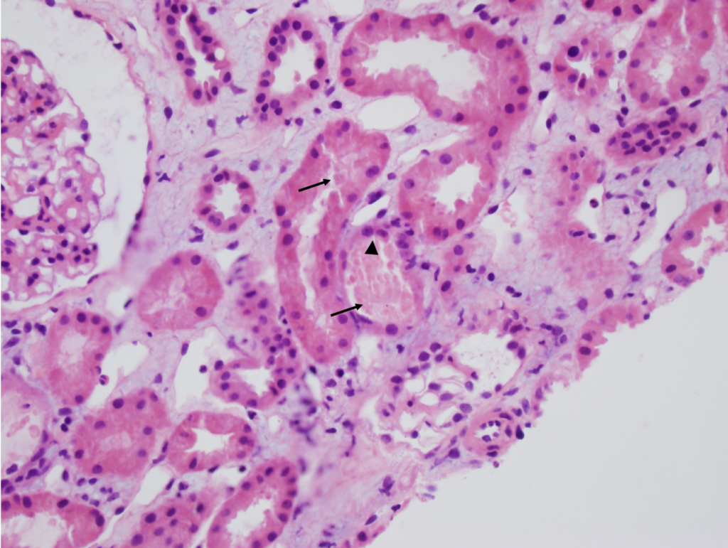
Numerous PAS-positive protein reabsorption droplets in the renal tubules of a child with minimal change disease. Image courtesy of Joseph Gaut, MD PhD.
Read MoreAcute tubular necrosis
Acute tubular necrosis (ATN). Note the tubules are not back-to-back due to interstitial edema (Masson trichrome staining, not shown, did not show appreciable fibrosis). There is blebbing and sloughing of…
Read MoreSLE nephritis
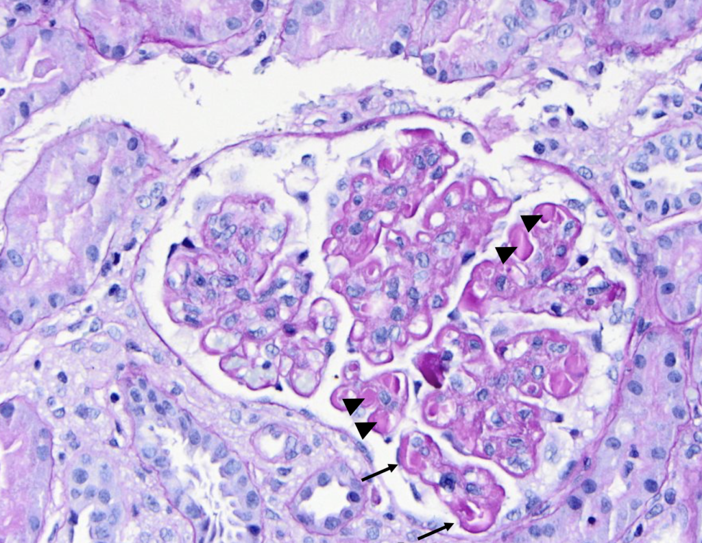
PAS stain of a glomerulus in a patient with SLE nephritis. There is endocapillary and mesangial proliferation, as evidenced by thickened, occlusive capillary loops and increased mesangial cellularity. Note the…
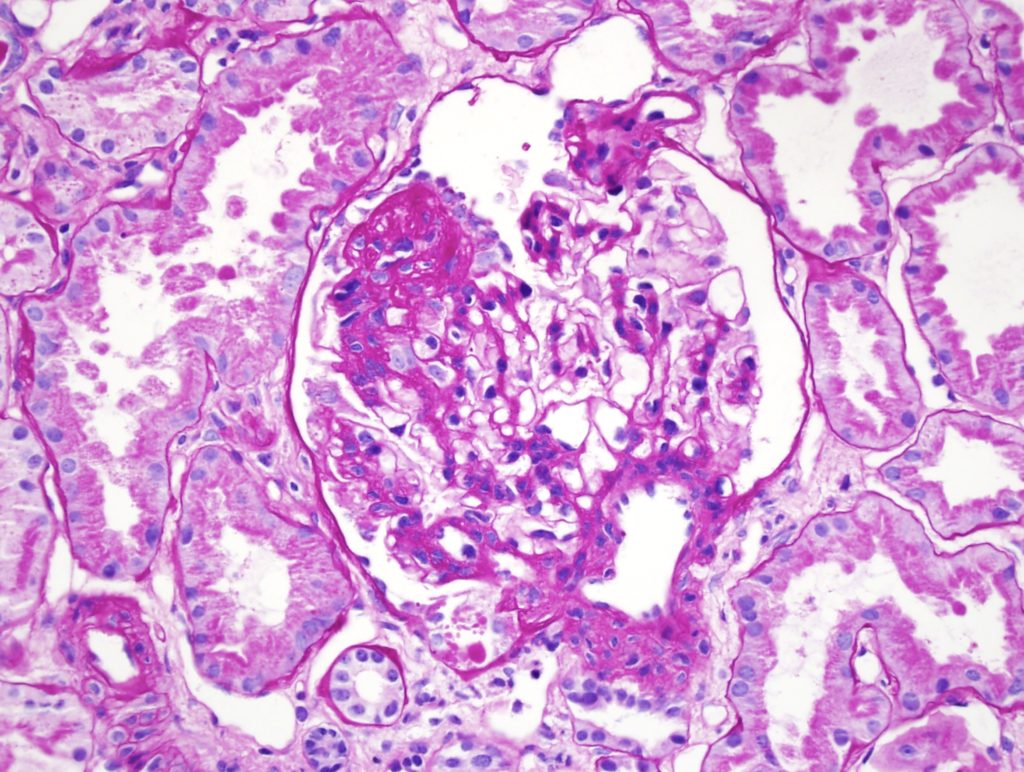
Read MoreFocal and segmental glomerulosclerosis
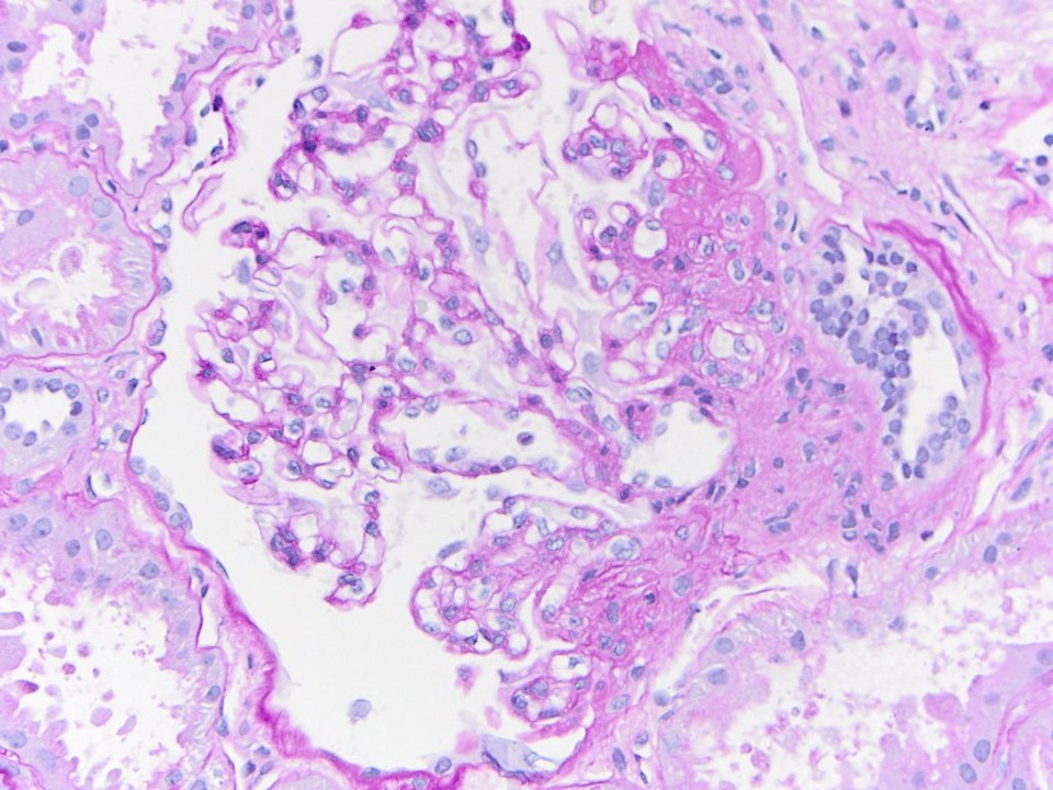
Segmental obliteration of the glomerular capillary lumen in a patient with FSGS. Note the sclerotic portion of the glomerular tuft is adherent to Bowman’s capsule. There is proximal tubular hypertrophy,…
Read More