Calyceal Diverticulum on MRU

Right anterior calyceal diverticulum on functional MR Urography. Top left side shows a T2 weighted image (no contrast) where fluid is bright, top right image is 10 min post-contrast…
Read MoreLarge neuroblastoma above left kidney CT scan coronal view
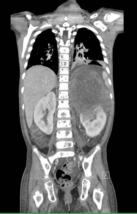
Large neuroblastoma mass above left kidney, distorting its architecture
Read MoreFocal and segmental glomerulosclerosis
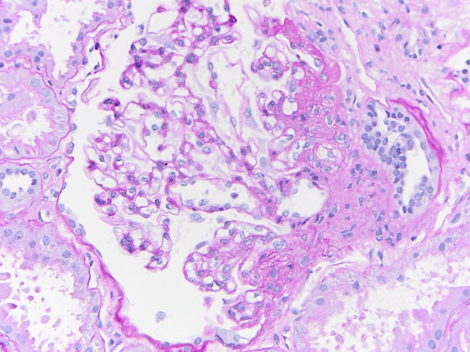
Segmental obliteration of the glomerular capillary lumen in a patient with FSGS. Note the sclerotic portion of the glomerular tuft is adherent to Bowman’s capsule. There is proximal tubular hypertrophy,…
Read More